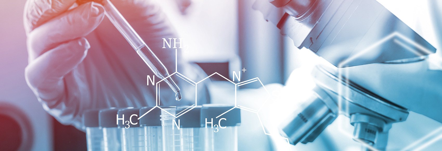
Immunohistochemistry (IHC) is an experimental technique based on the antigen-antibody reaction of immunology, which is used to analyze the metabolism, function and morphological changes of cells and tissues by applying physicochemical methods to display the material components of cell structures. Our company provides a one-stop IHC technology platform, which is widely used in medical research, especially in clinical tumor diagnosis and differential diagnosis.
Our IHC Technology Platform
IHC staining is the process by which antibodies recognize target antigens. This technology platform uses the principle of specific binding of antigens to antibodies to identify intracellular antigens (peptides and proteins) in tissues through a chemical reaction that causes color development of chromogenic agents (fluorescein, enzymes, metal ions, isotopes) labeled with antibodies for localization, characterization, and quantitative studies.
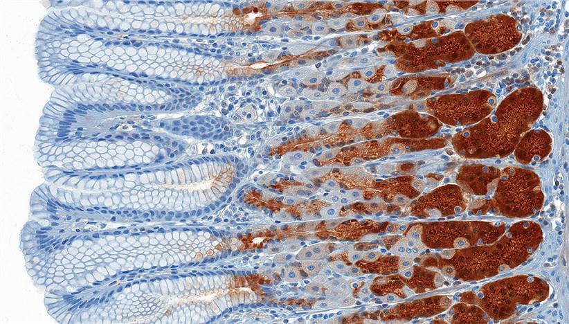
The antigen-antibody reaction can be seen by applying a color development assay. One method of color development involves the use of an enzyme-conjugated antibody as a substrate to produce pigmentation at the protein site. Another method is the fluorescence assay, in which a fluorescent dye is bound to the antibody and detected by fluorescence microscopy.
The main steps of our services are as follows
- Tissue processing, fixation, and sectioning.
- Antigen retrieval.
- Removal of endogenous peroxidase.
- Blocking.
- Primary and secondary antibody incubation.
- Detection.
- Counterstaining.
Tissue processing, fixation, sectioning
| Paraffin Sectioning | Frozen Sectioning |
| Fixed | Before embedding: formaldehyde | Before or after sectioning: formaldehyde, methanol, ethanol, or acetone |
| Slice | Slicer | Cryostat |
| Store | Store at room temperature for many years | One year at -80 ℃ |
Main methods of antigen retrieval
| Heat-induced epitope retrieval | Proteolytic enzyme-induced epitope retrieval |
| Temperature | About 95 ℃ | Typically 37 ℃ |
| Incubation time | 10-20 min | 10-15 min |
| pH | Typically, a pH 6 buffer is used, but alkaline buffers are also widely used. | The pH is usually 7.4 |
| Buffer Components | Commonly used buffers include sodium citrate, EDTA, and Tris-EDTA. | Neutral buffer for enzymes such as pepsin, proteinase K, or trypsin. |
Blocking
- Protein blocking: Bovine serum albumin (BSA) or casein can be used to block nonspecific antibody binding.
- Biotin Blocking: Incubate the tissue with avidin to block endogenous biotin, followed by incubation with exogenous biotin to block additional biotin binding sites on the avidin molecule.
Detection
- Enzyme Chromogenic Method: The most commonly used enzymes are HRP. Chromogenic detection is generally more sensitive than fluorescent detection. In addition, unlike fluorescent dyes, colored precipitates are photostable, so stained sections can be preserved for many years.
- Fluorescence method: Fluorescence detection is often used in situations where simultaneous detection of multiple antigens is required. Fluorescent dyes can be conjugated to primary or secondary antibodies or streptavidin.
IHC Service Description
What the customer provides
- Samples: Fixed tissues, tissue sections, cell crawls, embedded wax block tissues. For paraffin-embedded tissues, biopsy surgically excised specimens should be taken or fixed within 2 hours to avoid tissue autolysis and antigen denaturation, etc., affecting the immunohistochemical effect.
- Immunohistochemical reagents: In addition to primary antibodies and fluorescent secondary antibodies, the enzyme detection systems required to perform immunohistochemical detection tests are provided free of charge by Biotech. Primary antibodies and uncommon secondary antibodies are purchased by the customers themselves or by us on their behalf.
What Our company provides
Experimental steps, clear and reliable IHC result graphs, and result analysis reports.
Experimental Cycle
About 1-2 weeks.
For more information, please feel free to contact us.
Related Services
It should be noted that our service is only used for research, not for clinical use.